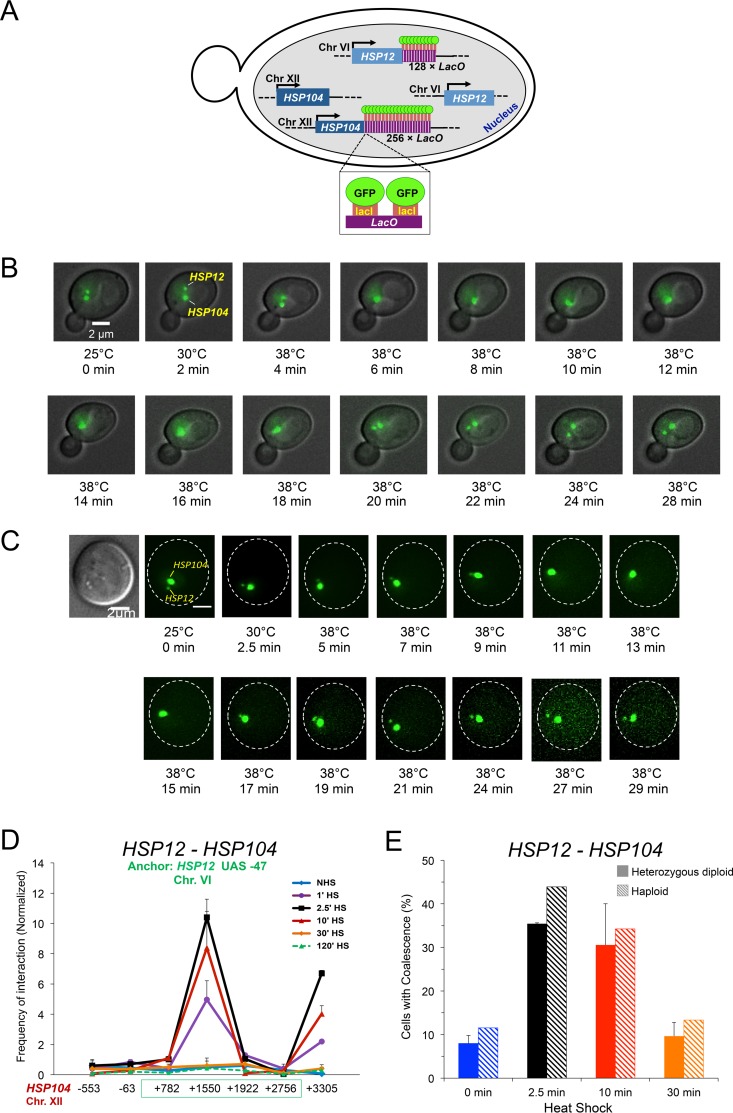
FIG 6.
Single-cell analysis demonstrates that HSP12 and HSP104 transiently coalesce in response to heat shock. (A) Schematic of ASK702, a diploid strain heterozygous for HSP12-lacO128 and HSP104-lacO256 and expressing LacI-GFP. (B) Live-cell fluorescence microscopy of an ASK702 diploid cell prior to and following application of heat for the times and temperatures indicated. The large and small green dots denote HSP104-lacO256 and HSP12-lacO128, respectively. Images were taken across nine planes in the z direction with an interplanar distance of 0.5 μm. Shown is a representative image for each time point. (C) Live-cell microscopy of an ASK702 cell responding to heat shock, as described for panel B. The first image was acquired using differential interference contrast (DIC). (D) TaqI-3C analysis of the kinetics of HSP12-HSP104 interaction, conducted as described for panel C. (E) Fraction of cells exhibiting HSP104-HSP12 coalescence. Mid-log-phase diploid and haploid cells (solid and hatched bars, respectively) were subjected to 30°C to 39°C heat shock for the indicated times and then fixed. For diploids (ASK706), 50 to 70 cells were evaluated per time point (means and SD are depicted; n = 2). For haploids (JTY001), 70 to 80 cells were evaluated per time point (n = 1).

