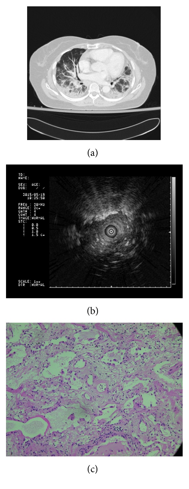
Figure 2.

A 66-year-old female received cryobiopsy from lingula showing correlation among the axial CT image, endobronchial ultrasound (EBUS) image, and histopathologic finding from biopsy. (a) Axial CT image in lung window demonstrated ground-glass opacities in peribronchial distribution and dependent atelectasis in the bilateral dependent lung. The image appearance is nonspecific with several possible differential diagnoses, including atypical pneumonia, acute interstitial pneumonitis, nonspecific interstitial pneumonitis, and/or pulmonary edema. (b) EBUS showed heterogeneous echogenicity, along with linear–discrete air bronchogram. (c) Histologic specimen of the biopsy revealed interstitial homogenous fibrosis and chronic inflammation (hematoxylin and eosin staining, 200x).
