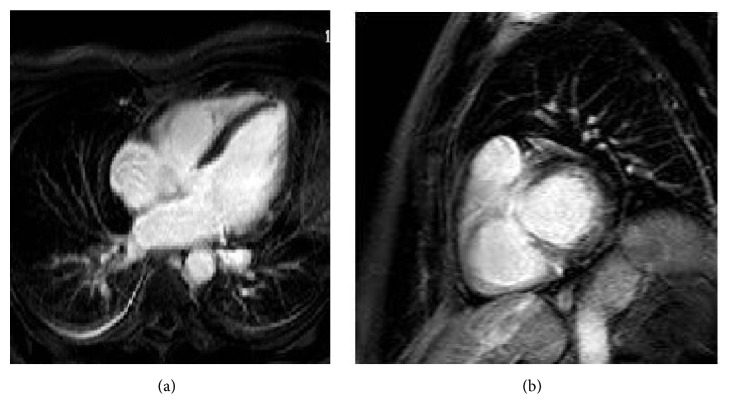
Figure 6.
LGE images of a patient with pulmonary sarcoidosis with cardiac involvement. Four-chamber view (a) showing mid-wall focal enhancement in the lateral wall of the left ventricle and in the apex. Note also the enhancement of the mediastinal lymph nodes. Short axis view (b) demonstrating enhancement of the LVOT and possibly of the RVOT. Images courtesy of Professor Dr. Jan Bogaert, University Hospital Leuven.

