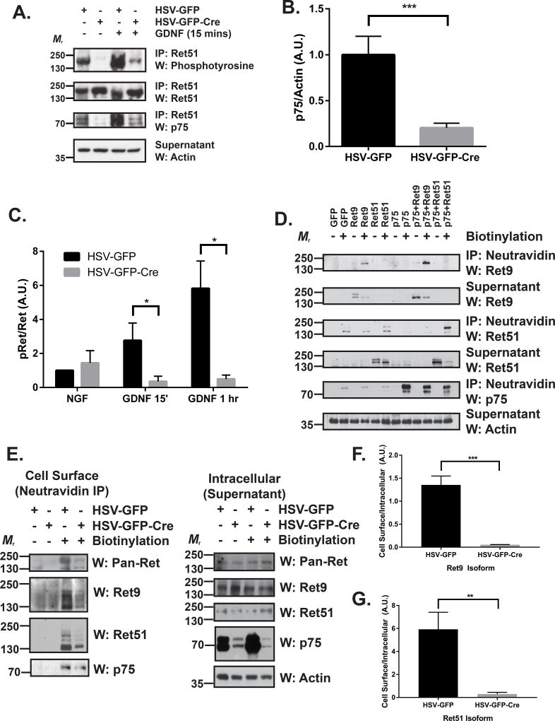
Figure 3. p75 augments GDNF-Ret signaling by increasing the cell surface localization of Ret.
(A) DRG neurons were cultured from P0 p75F/F mice in the presence of 50 ng/ml NGF. After two days, neurons were exposed to HSV-GFP or HSV-GFP-Cre viruses overnight (indicated above the blots). Neurons were maintained in culture until 7 DIV, followed by stimulation with medium alone or with 50 ng/ml GDNF for 15 minutes or 1 hour. Ret was immunoprecipitated and its level of activation was determined by phosphotyrosine immunoblotting. p75 immunoblotting confirmed that p75 protein levels were substantially reduced upon Cre expression. Actin immunoblotting of the supernatants served as a loading control. (B) Quantifications of p75 levels in 10 separate experiments demonstrate that approximately 80% of p75 is removed following treatment with HSV-GFP-Cre compared to HSV-GFP alone (*** p < 0.001). (C) Quantification of the mean proportion of phospho-Ret (pRet) over total Ret ± the standard error (5 experiments were quantified; * p < 0.05). (D) NIH/3T3 cells were transfected with GFP, Ret9, Ret51, p75, p75 and Ret9, or p75 and Ret51. After 24–36 hours, the cells were surface biotinylated using NHS-LC-Biotin in PBS (or treated with PBS alone as a control). The cells were then subjected to neutravidin precipitation. Precipitated cell surface proteins and supernatants (containing intracellular proteins) were then immunoblotted for Ret9, Ret51, and p75. Actin immunoblotting served as a loading control. (E) DRG neurons were cultured from P0 p75F/F mice and treated as described in A. Neurons were subsequently surface biotinylated as in D followed by neutravidin immunoprecipitation. Precipitated cell surface proteins (left) and supernatants (right; containing intracellular proteins) were analyzed by immunoblotting for pan-Ret (upper), Ret9 (2nd panel), and Ret51 (3rd panel). Actin immunoblotting served as a loading control. p75 immunoblotting confirmed efficient knockdown of greater than 70% of p75. (F) Quantifications of the cell surface levels of Ret9 (*** p < 0.001) and (G) Ret51 (** p < 0.01) indicate a highly significant difference in HSV-GFP-Cre treated neurons compared to HSV-GFP controls. IP, immunoprecipitation; W, western blot.

