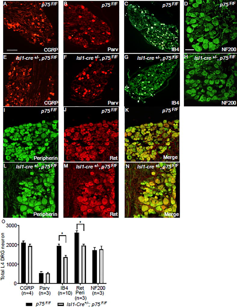
Figure 6. IB4+ nonpeptidergic nociceptors are selectively lost in Isl1-Cre+/−; p75F/F mice.
Immunofluorescence staining of adult L4 DRG with anti-CGRP (panel A and E), anti-Parvalbumin (panel B and F), anti-NF200 (panel D and H) antibody, IB4 (panel C and G) or a combination of anti-peripherin (I, L) and anti-Ret (J, M) antibodies from p75F/F (panel A–D, I–K) and Isl1-Cre+/−; p75F/F (panel E-H, L-N) mice. Scale bar for A-C, E-G, 200µm (located in panel A), Scale bar for D, H-N 100µm (located in panel D). (O) The quantification of CGRP+, Parvalbumin+, IB4+, Peripherin+/Ret+ and NF200+ neurons in adult L4 DRGs from p75F/F and Isl1-Cre+/−; p75F/F mice. Only the IB4+ and Peripherin+/Ret+ neurons were significantly reduced in the Isl1-Cre+/−; p75F/F mice. Values are expressed as mean ± SEM, * p<0.05. N indicates the number of animals being counted (each animal provides two DRGs).

