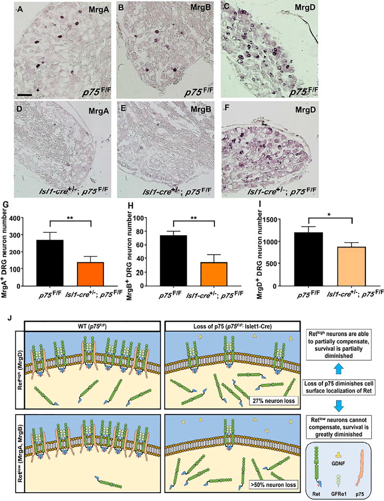
Figure 7. Nonpeptidergic neuron deficits in Isl1-Cre+/−; p75F/F mice correlates with level of p75-Ret co-expression.
In situ hybridization of adult (6 months old) L4 DRG with MrgA (panel A and D), MrgB (panel B and E) and MrgD (panel C and F) in situ probes from p75F/F mice (panel A–C) and Isl1-Cre+/−; p75F/F (panel D–F). Scale bar, 100µm. Quantification of MrgA+ (panel G), MrgB+ (panel H) and MrgD+ (panel I) neurons showed that in Isl1-Cre+/−; p75F/F mice, over 50% of MrgA+ and MrgB+ neurons were lost, while approximately 20% of MrgD+ neurons were lost. Importantly, MrgA+ and MrgB+ neurons were shown to express low level of Ret while MrgD+ neurons express high level of Ret. The value is expressed as mean ± SEM (n=3 for each data point, * p<0.05, ** p<0.01). (J) The expression of p75 increases the level of Ret on the cell surface in p75F/F mice and potentiates GFL-induced Ret phosphorylation and its downstream survival effects. When p75 expression is lost in Isl1-Cre+/−; p75F/F mice, the level of Ret on the cell surface is reduced. For neurons that express high levels of Ret (MrgD+), the high level of Ret expression may compensate for the loss of p75 and results in more modest deficits. In neurons that express low levels of Ret (MrgA+ and MrgB+), however, the loss of Ret on the cell surface cannot be compensated for, leading to substantial cell loss.

