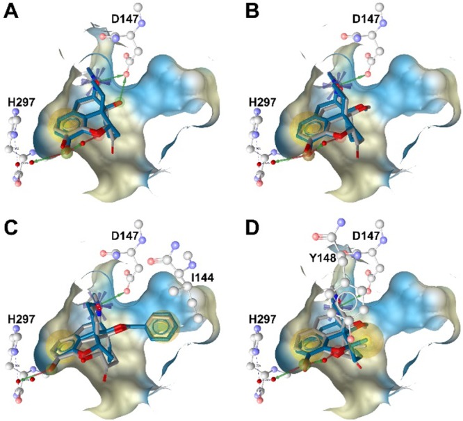
Figure 2.

Docking of N-methylmorphinans-6-ones 1-4 (gray) and corresponding 6-desoxo counterparts 1a–4a (blue) to the active structure of the MOR: (A) overlay of 1 and 1a; (B) overlay of 2 and 2a; (C) overlay of 3 and 3a; (D) overlay of 4 and 4a. The key residues D147 and H297, involved in a hydrogen-bonding network, and residues I144 and Y148, involved in hydrophobic interactions, are depicted. Chemical features are color-coded: red/green arrow, hydrogen bond acceptor/donor; yellow sphere, hydrophobic interaction; blue asterisk, positively ionizable. Binding pocket surface is shown, colored according to aggregated hydrophilicity/hydrophobicity (blue/ochre).
