Fig. 2.

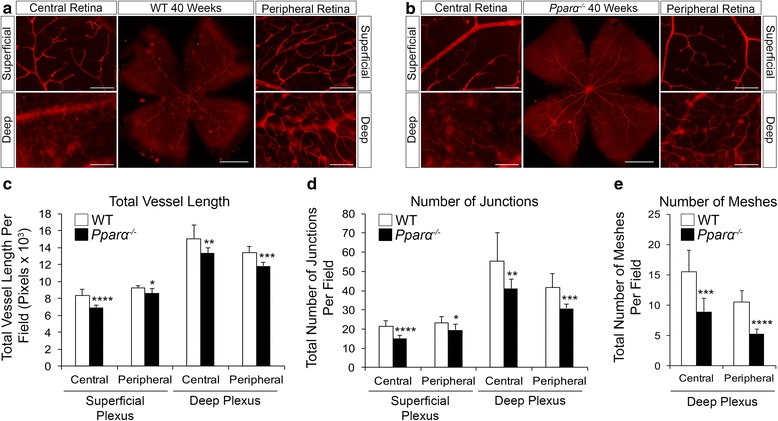
Decreased vascular density in Pparα -/- retinas. a Representative images of isolectin-labeled microvessels in retinal wholemount and single fields of superficial and deep vascular plexuses in central and peripheral retina of 40-week-old wild-type (WT) retinas. b Representative images of 40-week-old Pparα -/- retinas. c Decreased total vessel length in superficial and deep vascular plexuses of Pparα -/- retinas. d Decreased number of vascular junctions in superficial and deep vascular plexuses of Pparα -/- retinas. e Decreased number of meshes in deep vascular plexus of Pparα -/- retinas (adult retinas do not have meshing in the superficial plexus) (WT, n = 7; Pparα -/-, n = 8 retinas). *P ≤ 0.05; **P ≤ 0.01; ***P ≤ 0.001; ****P ≤ 0.0001, Student’s t test
