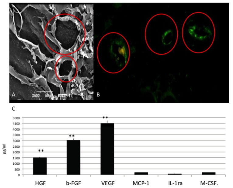
Figure 2.
(A) SEM(scanning electron microscope ) analyses of HUVEC cultured onto Osteon Growth Induction (OGI) surfaces; (B) immunoistochemical test against CD31 (in green), capillary like structures (red circles) as SEM analyses revealed; (C) Quantification of growth factor release pg/mL. Statistically significant differences are indicated as ** p < 0.01.

