Fig. 6.

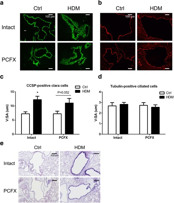
The immunofluorescent expression of epithelial cells (Clara cells, ciliated cells) and airway mucus. Examples of CCSP-positive Clara cells (green) and their group data are illustrated in (a) and (c) with the bar equal to 100 μm, while examples of Tubulin-positive ciliated cells (red) and their group data in (b) and (d). V:SA is the ratio of the volume to the surface area. In C, results are expressed as mean ± SEM. Numbers of mice were six in each group; * P < 0.05, HDMIntact versus CtrlIntact group. And P = 0.052, HDMPCFX versus CtrlPCFX group. e AB-PAS stain (bars = 200 μm). Images are representative micrographs of lung tissues obtained from each group
