
Fig. 4.
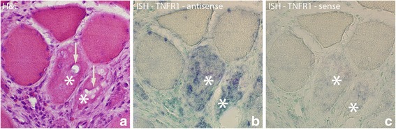
Parallel sections of a group of muscle fibers from 1 week group (experimental side) which are investigated with H&E, ISH antisense and sense TNFR1 mRNA probes. There are two muscle fibers (marked with *) that are different from the others surrounding them, and in which TNFR1 mRNA is present, exhibiting a diffuse but localized pattern (b). Note the presence of vacuoles (arrows, a) in these muscle fibers. Note also the frequently occurring white blood cells in the connective tissue outside the muscle fibers. That includes eosinophils (red coloured) (a). There is no specific reaction in the control (c). Orig. magnif. ×300