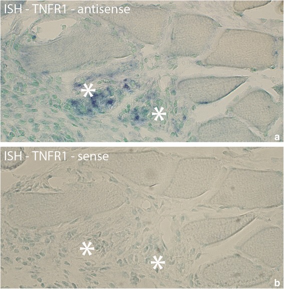
Fig. 5.

Sections from experimental animal (3 week group, experimental side) processed with ISH; antisense (a) and sense (b) probes. a shows that there are TNFR1 mRNA reactions in two muscle fibers (asterisks) that are very poorly outlined. b is a control (use of sense probe). In (b), as well as in (a), it can be noted that there is cellular infiltration in the two muscle fibers, especially the one to the right, suggesting that they are necrotic fibers. Both necrotic fibers are very much destroyed. Such an infiltration of cells was not seen in the muscle fibers expressing TNFR1 mRNA in Fig. 4. Note that the other muscle fibers seen in the fig appear normal. Orig. magnif. ×300
