
Fig. 3.
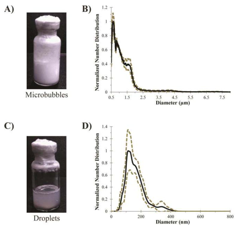
Microbubble condensation to form submicrometer droplets from volatile compounds (from [23]). (a) Phospholipid-encapsulated DFB microbubbles initially appear as an opaque “milky” emulsion. (b) Produce a distribution with a mean diameter of 1.0 ± 0.9 μm (N = 3, Accusizer 780, five-point smoothing applied). (c) After condensation, the emulsion appearance turns translucent. (d) Distribution shifts into the nanoscale with a mean diameter of 164 ± 63 μm (N = 3, Malvern NS-500). Dashed lines represent one standard deviation.