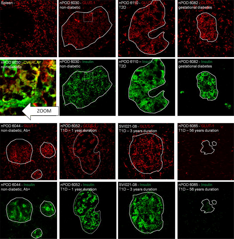
Figure 4.
Immunofluorescent glucose transporter 1 (GLUT1) expression survey on pancreatic islets from donors with various diabetic conditions. All representative samples are individually labelled with sample and staining ID. Spleen section from a normal individual served as a positive control for staining, while nPOD6065 in which no GLUT1 mRNA was detected is a negative control. From this staining series of it can be concluded that GLUT1 is the dominant isoform on human pancreatic islets and persist after many years of type 1 diabetes on remnant insulin-positive islets. The zoom shows yellow colocalization between insulin and GLUT3, indicating that beta cells are among the cells that express this transporter. White outlines demarcate insulin-positive areas and were superposed onto the GLUT1 images for comparison. White squares are zoom regions

