
FIGURE 7.
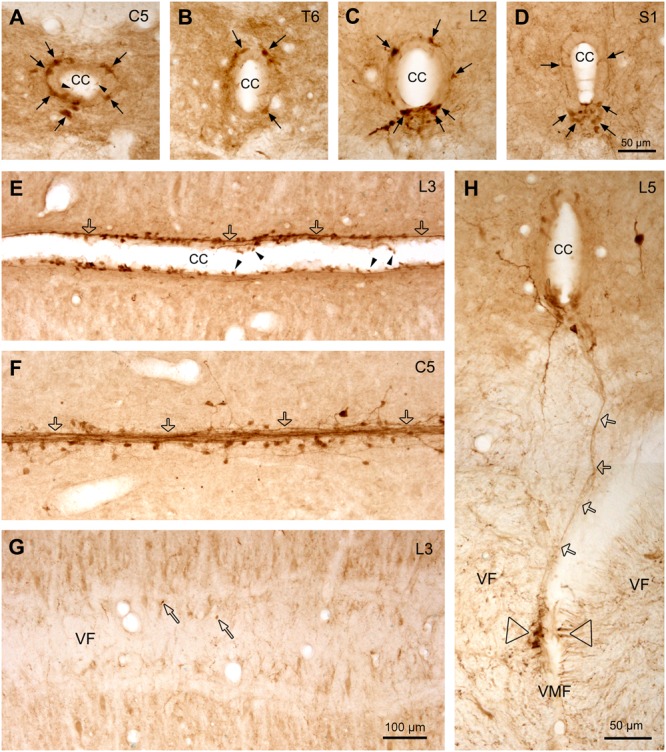
Aromatic L-amino acid decarboxylase cells and fibers/fiber bundles around the CC. (A–D) AADC cells around the CC from C (A), T (B), L (C), and S (D) segments illustrated in transverse sections. Dorsal side is upward for all the sections. It is clear that at C and T spinal levels the AADC cells were distributed evenly around the CC in different directions, whereas at the L and S levels, especially at S level, the AADC cells were mainly located ventral to the CC although a small number of the cells could also be observed in other parts (arrows). (E,F) AADC cells and fibers/fiber bundles lateral to the CC (E) and just at the bottom of the CC (F) seen in horizontal sections. It seems that the fibers arising from the AADC cells joined together to form fiber bundles (hollow arrows) which run longitudinally along the CC, yielding garlic braid-like chains. (G) Image from a horizontal section just below the CC showing that only a few AADC-IR puncta are apparent (narrow hollow arrows) in VF, indicating that most fibers from the AADC cells around the CC do not project toward the VMF. (H) Image from a transverse section showing a AADC-IR fiber/fiber bundle (hollow arrows) ran all the way from the ventral side of the CC to the left side of the VMF, where a cluster of AADC-immunoreactive fibers is apparent (hollow triangles). This fiber/bundle might be originated from an AADC cell(s) around the CC but it is hard to pinpoint its originating cell body in this section. In some sections, e.g., in (A) and (E), clear swellings at one end of the AADC cell processes could be seen to protrude into the lumen of the CC (arrowheads). The spinal segment level is indicated at upper right corner in each panel. AADC antibody was from rabbit. Scale bar in (D), valid for (A–D), and in (H), 50 μm; in (G), valid for (E–G), 100 μm.