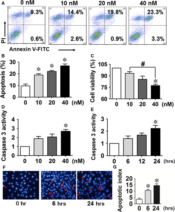
Figure 1.

2, 3, 7, 8‐tetrachlorodibenzo‐p‐dioxin (TCDD) exposure increases apoptosis in human umbilical vein endothelial cells (HUVECs). (A) Annexin V‐FITC/PI staining of HUVECs treated with different concentrations of TCDD for 24 hrs was assessed by flow cytometry. The percentages of early and late apoptotic cells are presented in the lower right and upper right quadrants, respectively. (B) Columns represent the average proportions of apoptotic cells. *P < 0.05 versus the 0 nM group; n = 3. (C) Cell viability in HUVECs exposed to 0, 10, 20 and 40 nM TCDD was analysed by the CCK‐8 assay kit. *P < 0.05 versus the 0 nM group, # P < 0.05 versus the 10 nM group; n = 3. (D–E) The effects of various TCDD doses (0–40 nM) and duration of exposure (at 40 nM) on caspase 3 activity in HUVECs. *P < 0.05 versus the 0 nM group; n = 3. (F) Apoptotic morphology of HUVECs incubated with 40 nM TCDD for 6 or 24 hrs after DAPI staining. Apoptotic cells were identified by condensation and fragmentation of nuclei. (G) The apoptotic index (AI) was calculated as the percentage of apoptotic nuclei per total nuclei number per field. Values represent mean ± SEM of four independent experiments, *P < 0.05 versus the 0 nM group; n = 4.
