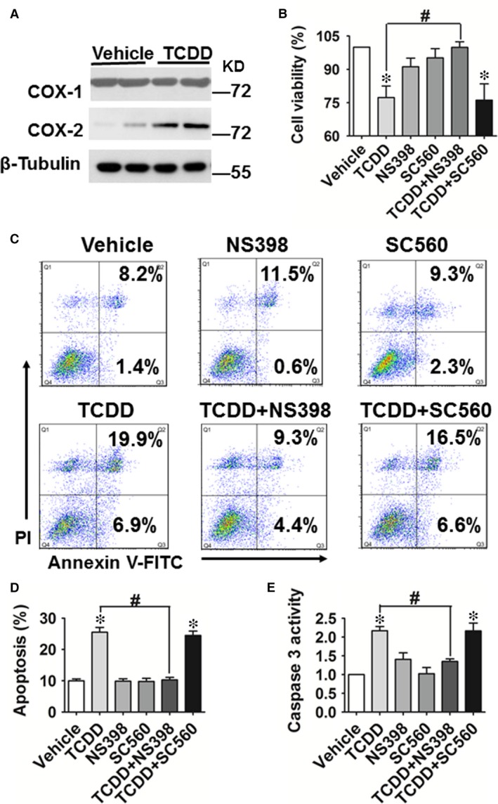
Figure 2.

COX‐2 is involved in 2, 3, 7, 8‐tetrachlorodibenzo‐p‐dioxin (TCDD)‐caused apoptosis in human umbilical vein endothelial cells (HUVECs). (A) Protein levels of COX‐1 and COX‐2 in HUVECs treated with or without TCDD (40 nM) for 24 hrs were analysed by Western blot. (B) Cell viability in HUVECs incubated with TCDD (40 nM), the COX‐2 inhibitor, NS‐398 (20 μM), the COX‐1 inhibitor, SC‐560 (20 μM), TCDD + NS‐398 or TCDD + SC‐560. *P < 0.05 versus vehicle, # P < 0.05 versus TCDD; n = 3. (C) Annexin V‐FITC/PI staining of HUVECs treated with TCDD (40 nM), NS‐398 (20 μM), SC‐560 (20 μM), TCDD + NS‐398 or TCDD + SC‐560 was assessed by flow cytometry. The percentages of early or late apoptotic cells are presented in the lower right and upper right quadrants, respectively. (D) Columns represent the proportions of apoptotic cells. *P < 0.05 versus vehicle, # P < 0.05 versus TCDD; n = 8–9. (E) Caspase 3 activity in HUVECs was measured using a Caspase 3 Activity Assay Kit. The treatments of HUVECs were the same as those described in Figure 1. *P < 0.05 versus vehicle, # P < 0.05 versus TCDD; n = 3.
