Figure 8.

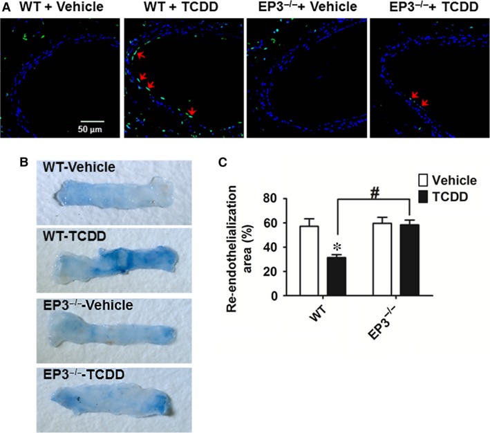
EP3 deficiency protects against 2, 3, 7, 8‐tetrachlorodibenzo‐p‐dioxin (TCDD)‐caused endothelial cell apoptosis in mice. Femoral artery injuries were induced by wire insertion in EP3−/− and WT mice, TCDD (10 μg/kg body weight) was injected to mice intraperitoneally every other day, and the injured femoral arteries were collected on day 10 for TUNEL assay and Evans blue dye staining. (A) Representative confocal microscope images of TUNEL‐stained femoral arteries. Blue indicates DAPI staining, and green indicates TUNEL staining. Arrows indicate TUNEL‐positive cells. Scale bar, 50 μm. (B) Representative photomicrographs of femoral arteries stained by Evans blue dye on day 10 after injury. The area stained blue corresponds to the area not yet re‐endothelialized. (C) Quantification of the re‐endothelialized area assessed by percentage of the area unstained with Evans blue dye in the entire injured area. *P < 0.05 versus WT + vehicle; n = 9–10.
