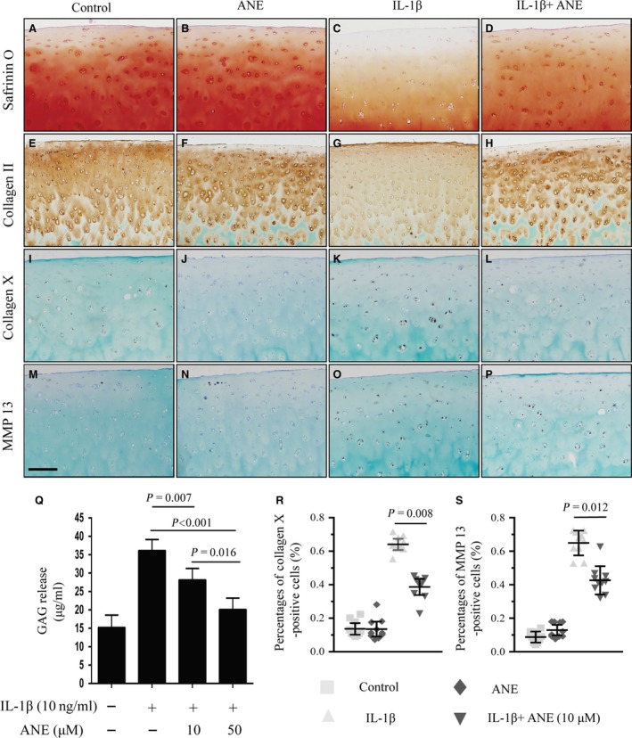
Figure 4.

Effects of anemonin on human articular cartilage ex vivo. The full‐thickness human cartilage explants were obtained from adult human joint tissues (n = 10, Mankin score 0–2). Cartilage explants were cultured in the absence or presence of IL‐1β (10 ng/ml) and anemonin (10 μM) for 4 days. (A–D) 5‐μm paraffin sections were stained with Safranin O–fast green to examine the cartilage proteoglycan loss. (E–H) Immunohistochemical staining for collagen II, (I–L) collagen X and (M–P) MMP13 in human articular cartilage. (R) Collagen X‐ and (S) MMP13‐positive cells in human articular cartilage were counted. (Q) Culture medium were collected, and the amount of GAG release into the medium was quantified by DMMB assay. GAG released into the medium was normalized as mass of GAG per millilitre (ml) of culture medium. Scale bar: 200 μm (A–P). Data are expressed as the mean (symbols) ± 95% confidence intervals (error bar).
