
Figure 1.
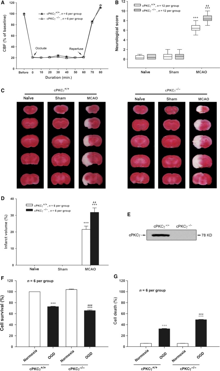
Effects of cPKCγ on regional CBF, neurological score and infarct volume of mice after 1‐hr MCAO/24‐hr R, and cell survival and death rates of cultured cortical neurons after 1‐hr OGD/24‐hr R. (A) The change in CBF of mice with MCAO treatment (n = 6 per group). (B) Statistical results of neurological score (n = 12 per group). Representative photographs of TTC‐stained coronal brain sections (C) and statistical results of infarct volume (D) from Naïve, Sham and MCAO groups of cPKCγ+/+ and cPKCγ−/− mice (n = 6 per group), respectively. (E) Representative results of Western blot demonstrated that cPKCγ was completely knocked out in mice. Quantitative analysis of the cell survival rate (F) and the cell death rate (G) from Normoxia and OGD groups of cPKCγ+/+ and cPKCγ−/− cortical neurons (n = 6 per group), respectively. +++ P < 0.001 versus Sham of cPKCγ+/+ mice; && P < 0.01 versus corresponding cPKCγ+/+ mice with the same treatment; ***P < 0.001 versus Normoxia of cPKCγ+/+ neurons; ### P < 0.001 versus corresponding cPKCγ+/+ cortical neurons with the same treatment.