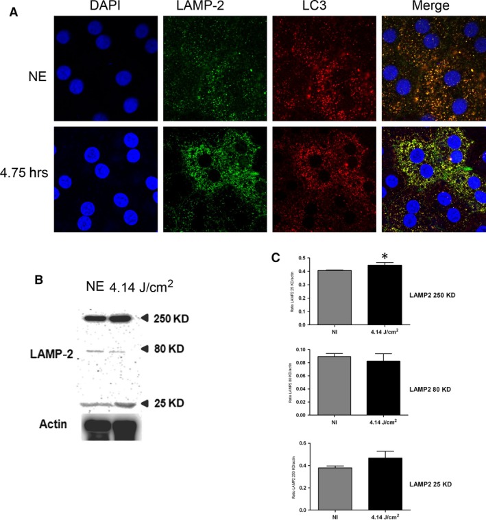
Figure 6.

Physiological function of autophagy after LED exposure. Male Wistar rats aged 7 weeks (n = 4) were exposed to white LED for 4.75 hrs. NE: Non‐exposed rats. (A) At the end of the exposure period, the RPE was flat‐mounted and immunostained with two combinations of antibodies: anti‐LC3 in red and with anti‐LAMP2 in green. The RPE was further analysed by triple staining with DAPI (blue) by confocal microscopy. The pictures were taken on the upper retina 100 μm away from the optic nerve. Scale bar represents 20 μm. (B) After LED exposure, the eyes were enucleated, the RPE dissected, extracted with M‐PER buffer and loaded on the top of a 10% SDS–PAGE, transferred onto a nitrocellulose membrane and probed for anti‐LAMP2. Actin was used as a charge control. (C) The histogram shows a quantification of p62 expressed as a ratio to actin * =P < 0.05).
