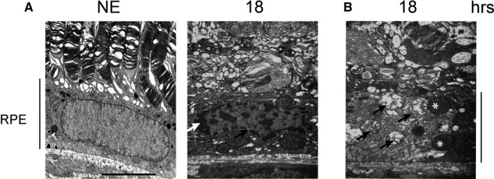
Figure 10.

Structural modifications of the RPE examined by transmittance electron microscopy (TEM). Male Wistar rats aged 7 weeks (n = 4) were exposed to white LED for 18 hrs (15.7 J/cm2). At the end of the exposure period, the eyes were analysed by TEM. NE: Non‐exposed animals. (A) After light exposure, the RPE presents peripheral condensation of chromatin (white arrow) and dilated nuclear pores (black arrow). (B) LED exposure leads to an alteration of the mitochondria. The black arrows point highly damaged mitochondria. The white stars indicate condensed mitochondria. The pictures were taken on the upper retina 100 μm away from the optic nerve. Scale bar represents 5 μm. RPE: retinal pigment epithelium.
