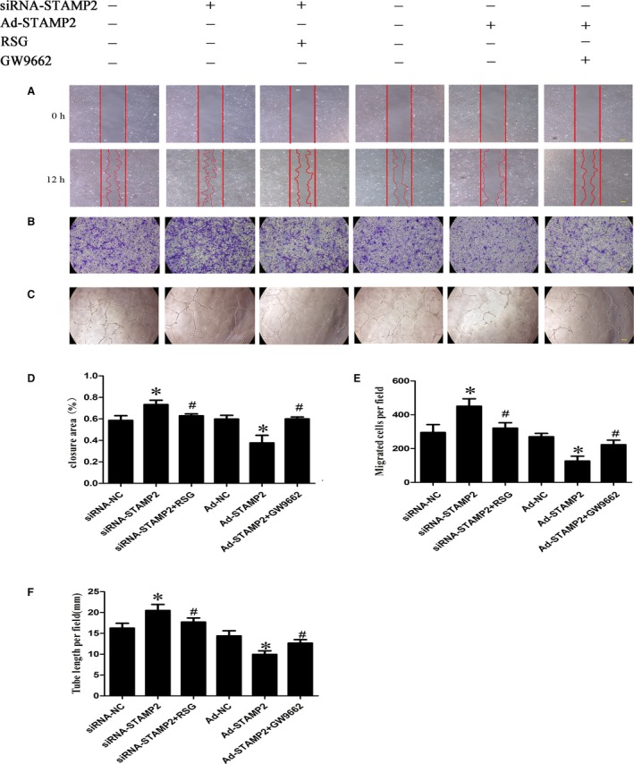
Figure 4.

Effect of STAMP2 on HUVECs migration and tube formation in vitro (n = 3 per group). (A) Representative images of wound‐healing assay at 0 and 12 hrs, respectively, showing that STAMP2 silencing increased human umbilical vein endothelial cell migration, whereas STAMP2 overexpression inhibited human umbilical vein endothelial cell migration. After activation or inhibition PPARγ by RSG or GW9662, the effects of STAMP2 silencing or STAMP2 overexpression on HUVECs are reversed. (scale bar: 100 μm); (B) Quantitative analysis of wound‐healing assay in six groups of human umbilical vein endothelial cells at 12 hrs. (C) Representative images of Transwell assay showing that STAMP2 silencing increased human umbilical vein endothelial cell migration, whereas STAMP2 overexpression inhibited human umbilical vein endothelial cell migration. After activation or inhibition PPARγ by RSG or GW9662, the effects of STAMP2 silencing or STAMP2 overexpression on HUVECs are reversed. (scale bar: 100 μm); (D) Quantitative analysis of transwell assay in six groups of human umbilical vein endothelial cells. (E) Representative images of tube formation assay showing STAMP2 silencing increased human umbilical vein endothelial tube formation, whereas STAMP2 overexpression inhibited human umbilical vein endothelial tube formation. After activation or inhibition PPARγ by RSG or GW9662, the effects of STAMP2 silencing or STAMP2 overexpression on HUVECs are reversed. (scale bar: 100 μm). (F) Quantitative analysis of tube formation assay in six groups of human umbilical vein endothelial cells. Data are mean ± SD; *P < 0.05 versus siRNA‐NC group/Ad‐NC group; # P < 0.05 versus siRNA‐STAMP2 group/Ad‐STAMP2 group.
