
Figure 6.
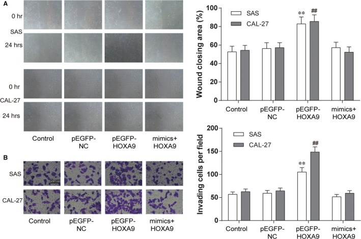
MiR‐139‐5p attenuated migration and invasion of OSCC cells by targeting HOXA9. (A) At 48 hrs after transfection, cell migration was assessed by wound healing assay. (B) Cell invasion was analysed by Transwell assay. Data are expressed as mean ± S.D. **P < 0.01 compared with control in SAS cell line, ## P < 0.01 compared with control in CAL‐27 cell line. Control: control group, cells were without transfection. pEGFP‐NC: pEGFP‐NC group, cells transfected with empty pEGFP plasmids. pEGFP‐HOXA9: pEGFP‐HOXA9 group, cells transfected with pEGFP‐N1‐3FLAG‐HOXA9‐GFP plasmids. MiR‐139‐5p mimics + pEGFP‐HOXA9: miR‐139‐5p mimics + pEGFP‐HOXA9 group, cells cotransfected with miR‐139‐5p mimics and pEGFP‐HOXA9.