
Figure 3.
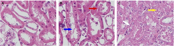
Renal lesions evaluated in the DCD model. Formalin‐fixed tissue stained with PAS. Panel A. Tubular epithelial cell flattening (TF). Panel B. Blue arrow indicates brush border loss (BBL), and red arrow indicates bleb formation (BF). Panel C. Yellow arrow indicates tubular lumen obstruction (TO) and white arrow tubular necrosis (TN). Magnification ×400.