
Figure 4.
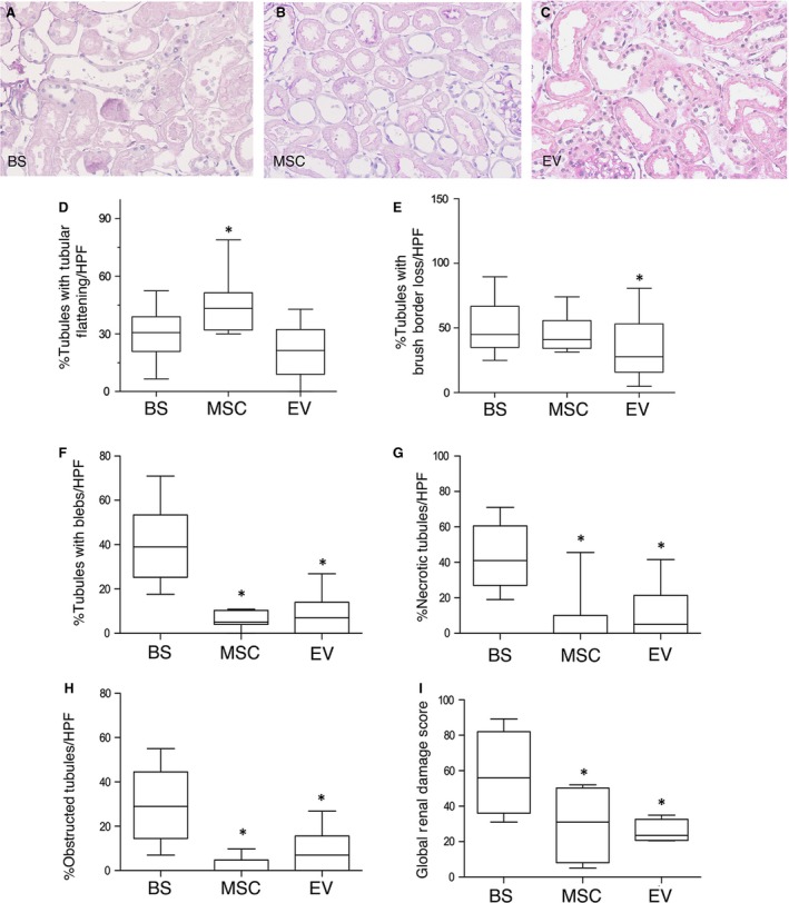
Ischaemic renal damage in BS and MSC‐/EV‐perfused kidneys. Representative renal sections of kidneys perfused after 20 min. of ischaemia either with Belzer solution (BS) (panel A), or Belzer solution supplemented with 3 million MSC (MSC) (panel B) or Belzer solution supplemented with EV derived from 3 million MSC (EV) (panel C). PAS staining, magnification ×200. Panels D‐I. Boxplots showing the distribution of renal lesions in all groups. Box: median, 25–75° percentile; whiskers, 5–95° percentile. Data are the percentage of tubules/HPF in which the lesions were observed. Panel D. *P < 0.05 versus BS and EV. Panel E. *P < 0.05 versus BS. Panel F. *P < 0.0001 versus BS. Panel G. *P < 0.0001 versus BS. Panel H. *P < 0.0001 versus BS. Panel I. *P < 0.0001 versus BS. TF: tubular epithelial cell flattening, BBL: brush border loss, BF: bleb formation, TO: tubular lumen obstruction, TN: tubular necrosis