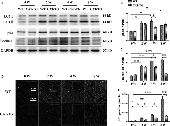
Figure 3.

Cardiac catalase overexpression ameliorates diabetes‐induced autophagy. Western blots showing protein levels of LC3‐II, p62 and beclin‐1, with GAPDH as a loading control (A). The band densities of p62 (B) and cleaved caspase‐3 (C) were semi‐quantitatively analysed. The presence of active autophagy was assessed in hearts by immunofluorescent detection of LC3‐II (D), and the LC3 dots were semi‐quantitatively analysed (E). LC3 fluorography magnification: ×60; bars = 50 μm. Values are mean ± S.E.M. Each group included six mice. *P<0.05, **P<0.01 and ***P<0.001.
