Figure 7.

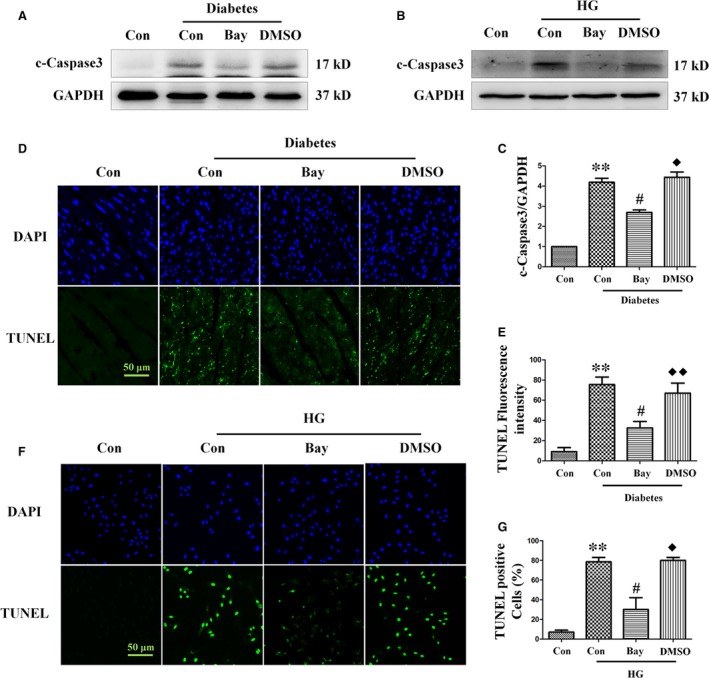
Treatment with the NF‐κB inhibitor Bay11‐7082 ameliorates diabetes‐induced apoptosis. Expression of cleaved caspase‐3 was assessed by Western blot analysis of diabetic heart tissue (A) and myocardial cells (B). Band densities were semi‐quantitatively analysed (C). The degree of apoptosis in heart tissue (D) and H9c2 cells (F) was assessed by TUNEL staining, followed by semi‐quantitative analysis (E and G, respectively). TUNEL magnification: ×60; bars = 50 μm. Values are mean ± S.E.M. Each group included six mice. In vivo results, **P<0.01 versus WT Control; #P<0.05, ##P<0.01 versus WT Diabetes; ◆P<0.05, ◆◆P<0.01 versus WT Diabetes+Bay11‐7082. In vitro results, **P<0.01 versus Control; #P<0.05 versus High glucose; ◆P<0.05 versus High glucose +Bay11‐7082.
