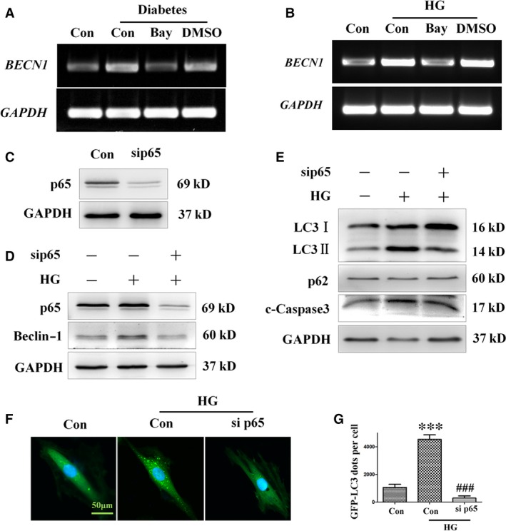
Figure 8.

Treatment with the NF‐κB inhibitor Bay11‐7082 ameliorates autophagy and apoptosis in H9c2 cells incubated in high‐glucose medium. H9c2 myocardial cells were pre‐treated with Bay11‐7082 or vehicle (DMSO) for 10 min. and then incubated in high‐ glucose medium (33 mM) for 24 hrs. RT‐PCR analysis of BECN1 expression in mice (A) and H9c2 cells (B). H9c2 myocardial cells were transfected with 40 nM p65 siRNA or 40 nM non‐specific (Data S1) control siRNA for 8 hrs and then incubated in 33 mM glucose medium for 24 hrs. Expression of p65 was assessed by Western blot analysis (C). The protein levels of p65, beclin‐1 (D), LC3‐II, p62 and cleaved caspase‐3 (E) were analysed by Western blot analysis. H9c2 cells infected with GFP‐LC3 adenovirus were transfected with 40 nM p65 siRNA or 40 nM non‐specific control siRNA for 8 hrs, and then incubated in high‐ glucose (33 mM) medium for 24 hrs. Autophagy in H9c2 cells transfected with siRNA targeting p65 was assessed by detection of GFP dots (F), which were semi‐quantitatively analysed (G). GFP‐LC3 fluorography magnification: ×60; bars = 50 μm. Values are mean ± S.E.M. Each group included six mice. ***P<0.001 versus Control; ###P<0.001 versus High glucose.
