
Figure 1.
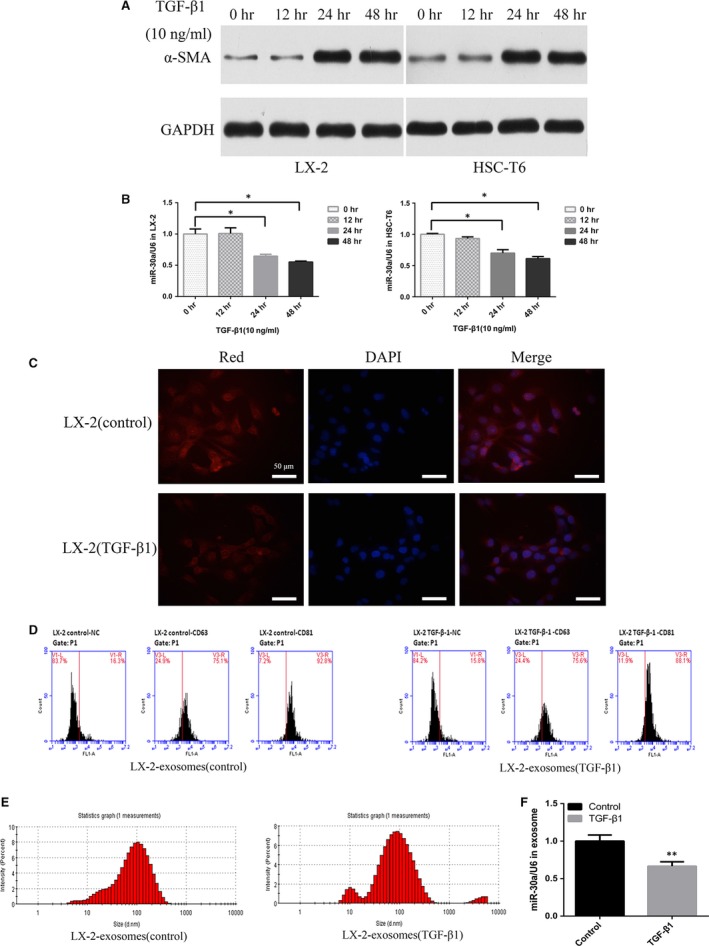
MiR‐30a is down‐regulated in activated HSCs and TGF‐β1‐treated LX‐2‐exosomes. HSCs were treated with TGF‐β1. (A) α‐SMA levels were detected by Western blotting. (B) miR‐30a levels were examined by TaqMan miRNA assay (*P < 0.05). (C) Detection of miR‐30a in LX‐2 cells using FISH, miR‐30a (Red) and nucleus (blue). (D) FCM analysis of surface markers (CD63 and CD81) on LX‐2‐exosomes. (E) Size detection of LX‐2‐exosomes. (F) MiR‐30a levels in LX‐2‐exosomes were examined by TaqMan miRNA assay. (**P < 0.01 versus the control group).