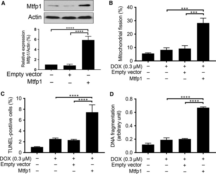
Figure 4.

Enforced expression of Mtfp1 sensitizes HL‐1 cells to doxorubicin‐induced mitochondrial fission and apoptosis. (A) Analysis of Mtfp1 expression. Immunoblot shows Mtfp1 overexpression in HL‐1 cells (upper panel). β‐actin served as a loading control. The densitometry data were expressed as the mean ± SEM of three independent experiments (lower panel). (B) Enforced expression of Mtfp1 sensitizes cells to undergo DOX‐induced mitochondrial fission. HL‐1 cells were exposed to a lower concentration of DOX. B shows percentage of cells with mitochondrial fission; (C and D) Enforced expression of Mtfp1 sensitizes cells to undergo DOX‐induced apoptosis. Cells were exposed to a lower concentration (0.3 μmol/l), and percentages of apoptosis were analysed by TUNEL assay (C) and DNA fragments were analysed using the cell death detection ELISA (D). Data were expressed as the mean ± SEM of three independent experiments. Figures presented are the representative figures of at least three independent experiments. ***P < 0.001 and ****P < 0.0001.
