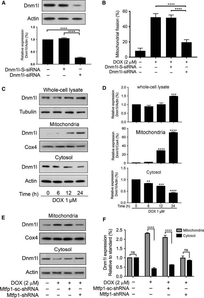
Figure 6.

Dnm1l is required for doxorubicin‐induced mitochondrial fission and Mtfp1 promotes doxorubicin‐induced Dnm1l accumulation in mitochondria. (A and B) Dnm1l is required for DOX to induce mitochondrial fission. (A) Analysis of Dnm1l expression. Immunoblot shows Dnm1l knockdown in HL‐1 cells. HL‐1 cells were transfected with scrambled siRNA, or Dnm1l siRNA, respectively, and after 48 hrs, were harvested for immunoblot (upper panel). Lower panel shows the densitometry. β‐actin served as a loading control. (B) Knockdown of Dnm1l inhibits doxorubicin (DOX)‐induced mitochondrial fission. B shows percentage of cells with mitochondrial fission. (C and D) DOX induces translocation of Dnm1l from cytosol to mitochondria. C shows Dnm1l expression by immunoblot. D shows the densitometry. **P < 0.01, ***P < 0.001 and ****P < 0.0001 versus 0 hr. (E and F) Knockdown of Mtfp1 inhibits DOX‐induced Dnm1l accumulation in mitochondria. Then, they were treated with a higher concentration (2 μmol/l) of DOX for 24 hrs and harvested for subcellular fraction, and protein expression levels in different cellular compartments were analysed by immunoblot. E shows Dnm1l expression in subcellular fraction and F shows densitometry. The densitometry data were expressed as the mean ± SEM of three independent experiments (lower panel). β‐actin served as a loading control for whole‐cell lysate and cytosolic component. Cox4 served as a loading control for mitochondrial component. Figures presented are the representative figures of at least three independent experiments. Data were expressed as the mean ± SEM of three independent experiments. ns, non‐significant; ****P < 0.0001.
