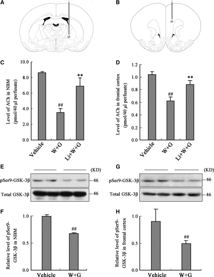
Figure 1.

Activation of GSK‐3 decreases ACh level in dialysates of NBM and frontal cortex. The microdialysis probes were implanted into the NBM and frontal cortex of the rats (A and B) at 30 min. after the left ventricular infusion of WT and GFX (W + G) or simultaneously combined LiCl (Li + W + G) or the vehicle. Then, the ACh level in the dialysates of NBM (C) and frontal cortex (D) was measured at 24 hrs after ventricular infusion. The Ser9‐phosphorylated GSK‐3β (pSer9‐GSK‐3β) and the total GSK‐3β were measured by Western blotting in NBM (E) and cortex (G) and relative quantitative analysis (F, H) at 24 hrs after the infusion. (LV, lateral ventricle; 3V, 3rd ventricle; D3V, dorsal 3rd ventricle; ## P < 0.01 versus Vehicle; **P < 0.01 versus W + G; n = 6).
