Figure 3.

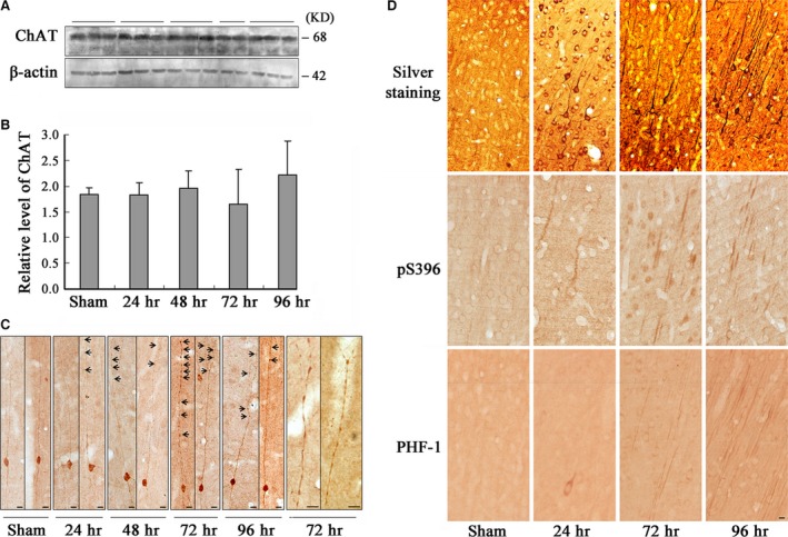
Activation of GSK‐3 causes axonal accumulation without affecting the level of ChAT in frontal cortex neurons. The rats were infused through left ventricle with WT and GFX; then, the frontal cortex slices and extracts were prepared at different time‐points as indicated (24–96 hrs). The level of ChAT was measured by Western blotting (A) and quantitative analyses normalized against β‐actin (B). Distribution of ChAT in the frontal cortex layer III pyramidal neurons was observed by immunohistochemistry, and the amplified images showing ChAT accumulation in axonal swellings at 72 hrs after infusion (C, arrowheads indicate axonal swellings). The enhanced argyrophil substances in the cell body and proximal neurites of the frontal cortex neurons at 24 hrs and the tangle‐like structures were shown at 72 and 96 hrs after infusion by Bielschowsky's silver staining (D, Silver staining). Similar pattern of the phosphorylated tau at Ser396 and PHF‐1 epitopes was shown by immunohistochemistry (D, pS396 and PHF‐1). (Bar = 10 μm; n = 6).
