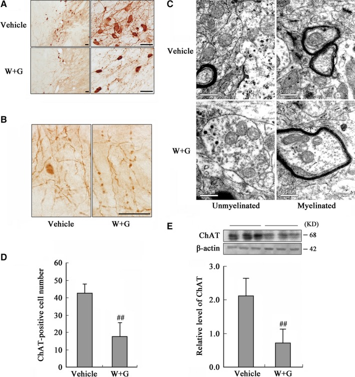
Figure 4.

Activation of GSK‐3 inhibits expression and causes accumulation of ChAT in NBM. The rats were infused through left ventricle with WT and GFX (W + G) or the vehicle for 24 hrs. (A) The distribution of ChAT in NBM neurons (Bar = 20 μm). (B) The amplified images showing axonal swellings (Bar = 20 μm). (C) Axon swelling measured by TEM (Bar = 0.5 μm). (D) The quantitation of ChAT‐positive neurons by stereological analysis. (E) The protein level of ChAT was measured by Western blotting, with β‐actin as an internal control. (## P < 0.01 versus Vehicle; n = 6).
