Figure 5.

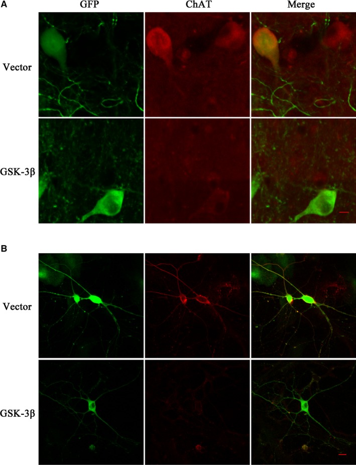
Overexpression of GSK‐3 inhibits ChAT protein level in NBM and primary cultured neurons of rat. We infected GSK‐3β AAV or vector virus in the rat NBM region for a month in vivo (A, green) and primary cultured neurons of the forebrain for 7 days in vitro (B, green), respectively. GSK‐3β with green fluorescence was detected by confocal laser scanning microscope. The red fluorescence represented ChAT protein in neurons. The pictures on the right column were the merged results of green and red fluorescence in relative groups. (Bar = 10 μm).
