Figure 9.

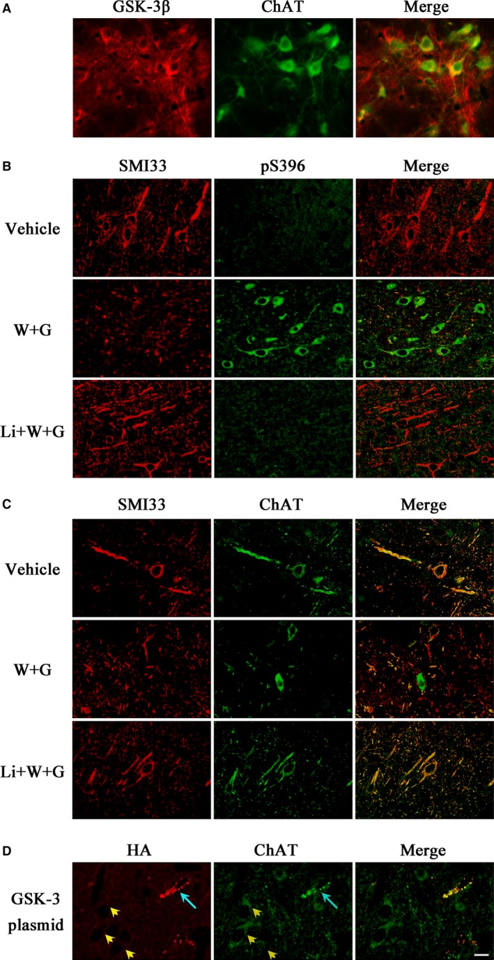
WT and GFX causes ChAT accumulation accompanied with the phosphorylation disorder of tau and neurofilaments in NBM. The simultaneous expression of GSK‐3β and ChAT was observed in NBM neurons of normal rat (A). Vehicle or WT and GFX (W + G) or simultaneously combined LiCl (Li + W + G) in a total volume of 2 μl was injected directly into NBM for 24 hrs, respectively. The expression and cellular distribution of phosphorylated tau (pS396), non‐phosphorylated neurofilaments (SMI33) and ChAT were measured by immunofluorescent staining (B and C). Wild GSK‐3β‐HA plasmid mixed with lipofectamine 2000 was injected into the NBM for 24 hrs, then the accumulation of ChAT was successfully detected in transfected cells (D, blue arrow), but not in the untransfected cells (yellow arrowhead). (Bar = 20 μm; n = 6).
