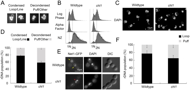
Fig 1. Chl1 helicase promotes rDNA condensation.
A) Representative examples of micrographs that highlight condensed (loop and line) and decondensed (amorphous puff-like and other non-discrete configuration) rDNA structures. B) Flow cytometer of DNA content at times indicated throughout the experimental procedure. Cells were maintained in nocodazole for 3 hours at 23°C post-alpha factor arrest. C) Chromosome mass and rDNA detected using DAPI in wildtype (YBS1019) and chl1 mutant (YBS1041) strains. D) Quantification of condensed (loop/line) and decondensed (puff/other) rDNA populations in wildtype and chl1 mutant cells. Data quantified from 3 biological replicates, 100 cells for each strain analyzed per replicate and statistical analysis performed using Student's T-test (p = 0.005). E) rDNA structures visualized using Net1-GFP, genome DNA detected using DAPI, and cell morphology images obtained using Differential Interference Contrast (DIC) microscopy. Yellow arrows indicate condensed rDNA loop/line and red arrowhead indicates decondensed rDNA puff. F) Quantification of condensed (loop) and decondensed (puff) rDNA populations in wildtype (YBS2020) and chl1 mutant (YBS2080) cells. Data quantified from 3 biological replicates, 100 cells for each strain analyzed per replicate and statistical analysis performed using Student's T-test (p = 0.006). Statistical significant differences (*) are based on p < 0.05.

