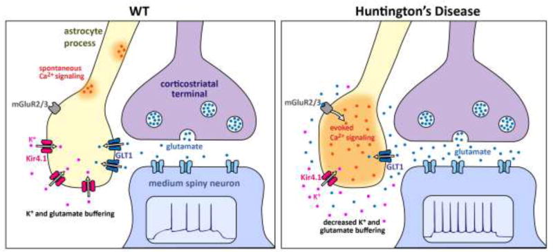
Figure 3. Schematic summary of the major Kir4.1, Ca2+ and GLT1 astrocyte changes observed in R6/2 mice at ~P70.
Only the key features are schematized and other differences between WT and HD mice are discussed in the text. In the WT striatum, normal expression levels of Kir4.1 and GLT1 maintain low levels of extracellular K+ and glutamate near synapses. As a result, electrical stimulation of cortico-striatal axons does not evoke robust mGluR2/3-mediated astrocyte Ca2+ signals. However, there are abundant spontaneous Ca2+ signals in striatal astrocytes. In HD mice, expression levels of Kir4.1 and GLT1 are reduced, and this has a number of observable consequences for astrocytes. Reduced GLT1 levels mean that astrocytes display robust mGluR2/3-mediated Ca2+ signals during stimulation of cortico-striatal axons. This gain of evoked Ca2+ signals is accompanied by a loss of spontaneous Ca2+ signals. The elevated glutamate and K+ levels in the extracellular space are proposed to contribute to increased excitability of MSNs. The cartoon is based on published work [33, 75, 81, 93].

