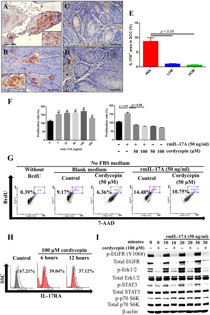
Figure 5. Both CMP and cordycepin inhibit IL-17A effects.
(A and B) Representative immunohistochemical staining of IL-17A in invasive squamous cell carcinoma (SCC) tissues of 4NA group are shown. Inserted pictures are of a higher magnification. (C and D) Immunoreactivity of IL-17A in invasive SCC tissues from LCM (C) and HCM (D) groups. Scale bar = 100 μm. (E) Quantification of IL-17A levels in SCC tissues of each group. Data are presented as mean ± SEM and statistical analysis is conducted with one-way ANOVA following Tukey’s test. (F) Recombinant IL-17A treatment for 72 hours stimulated proliferation of 4NAOC-1 cells, and cordycepin partially inhibited the proliferation. Data are presented as mean ± SD and statistical analysis is conducted with one-way ANOVA following Tukey’s test. Representative data from three independent experiments are shown. (G) Recombinant IL-17A treatment for 72 hours stimulated DNA synthesis in 4NAOC-1 cells and cordycepin partially inhibited the effect. Representative results are shown. BrdU (bromodeoxyuridine) was added at the last 24 hours of cultivation. (H) Cordycepin treatment for 6 and 12 hours decreased IL-17RA expression in 4NAOC-1 cells. Representative data are shown. (I) Cordycepin pre-treatment for 12 hours inhibited IL-17A/ IL-17RA signaling in 4NAOC-1 cells. Representative data from three independent tests are shown.

