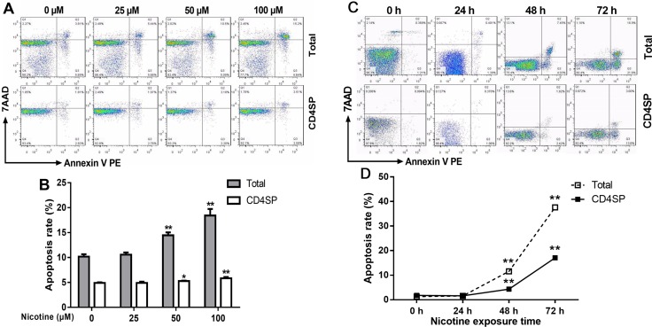
Figure 4. Effect of nicotine treatment in vitro on the apoptosis of primary thymocytes.
Thymus lobes dissected from infant mice at 3 weeks of age were gently dissociated over the stainless steel sieve. The cells were treated with different concentrations (25, 50 and 100 μM) of nicotine for 48 hours or 50 μM of nicotine for 24 h, 48 h and 72 h. The apoptosis frequency was determined by flow cytometry. (A, C) Typical flow diagram of thymocyte apoptosis; (B) Effects of different concentrations of nicotine (0–100 μM) on thymocyte apoptosis; (D) Effects of 50 μM nicotine exposure for different time (0, 24, 48, 72 h) on thymocyte apoptosis. The difference was analyzed with t-test. Mean ± SD, n = 3–4. *P < 0.05, **P < 0.01 vs control.

