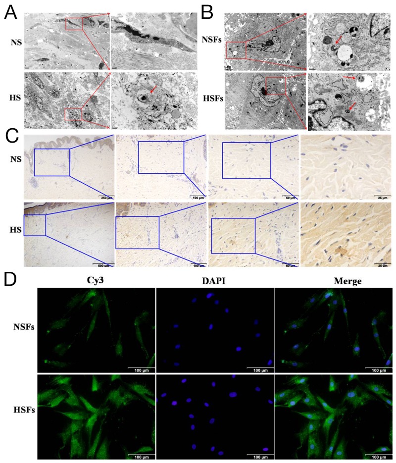
Figure 1. Autophagic capacity was elevated in HS/HSFs.
(A) Tissues were fixed and prepared samples by conventional TEM for observation. There was more autophagosomes in HS (red arrow) than in NS. Scale bars, 2 μm, 0.5 μm. (B) Cells were digested, washed, fixed and prepared samples. There were autophagosomes in HSFs similar to in NSFs. Scale bars, 2 μm, 0.5 μm. (C) Streptavidin-peroxidase DAB staining showed that LC3 was localized in HS tissue and NS tissue. LC3 was distributed in the cytoplasm, with more intensive staining in HS than in NS tissue. Scale bars, 250 μm, 100 μm, 50 μm, 25 μm. (D) NSFs and HSFs were grown on coverslips until they reached 70-80% confluence, fixed in 10% formaldehyde, washed, permeabilized, and blocked. Cells were incubated with an LC3B monoclonal antibody, followed by incubation with a corresponding Cy3-conjugated secondary antibody. The nuclei of the fibroblasts were stained with DAPI. Scale bars, 100 μm.

