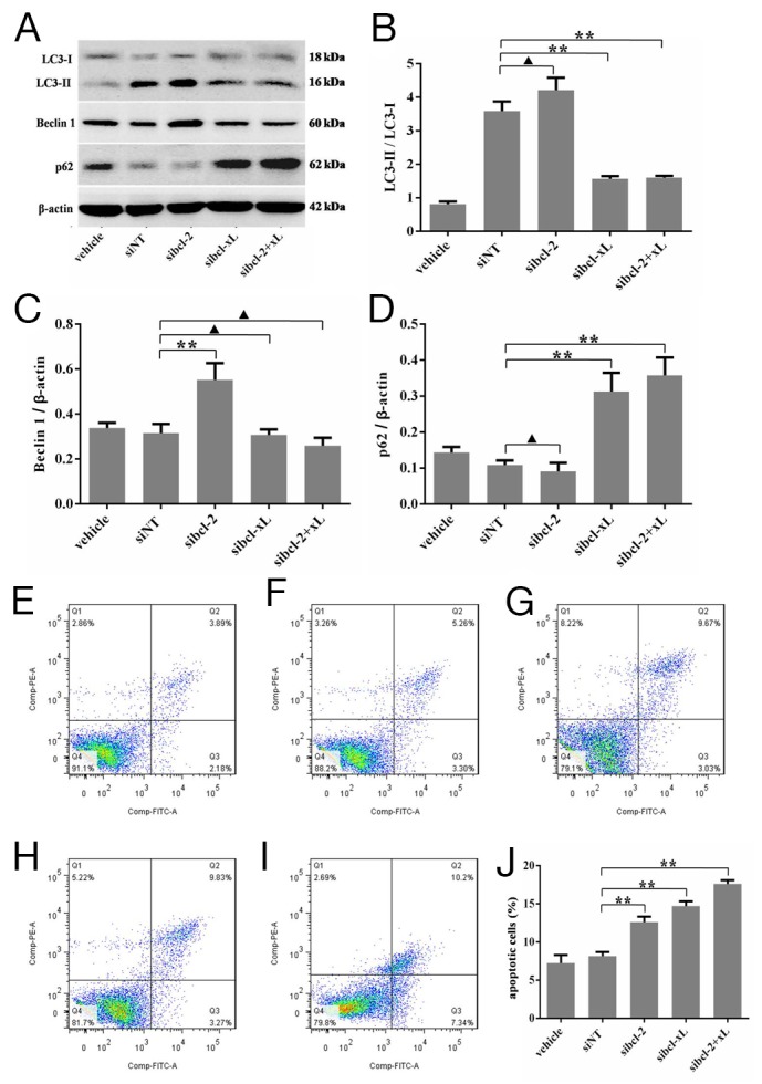
Figure 5. Silencing for Bcl-xL inhibited the autophagic capacity in HSFs, and promoted the apoptosis of HSFs.

HSFs, with 70-80% confluent, were transfected by siRNAs and their negative control, cultured in DMEM medium for 48 h, then Western blot analyses. (A, B) The ratio of LC3-II/LC3-L changes in transfected HSFs. (A, C) Autophagy protein Beclin 1 expresses and changes in the Beclin 1/β-actin ratio. (A, D) p62 protein expresses and changes in the p62/β-actin ratio. (E-J) HSFs, with 70-80% confluent, were transfected by siRNAs and their negative control, cultured in DMEM medium for 48 h. Apoptosis was measured by flow cytometric analysis of Annexin V and propidium iodide staining. Blank cells were used as control. Data are representative of three experiments. n = 3, ▲p > 0.05, *p < 0.05, **p < 0.01 compared with the negative control group.
