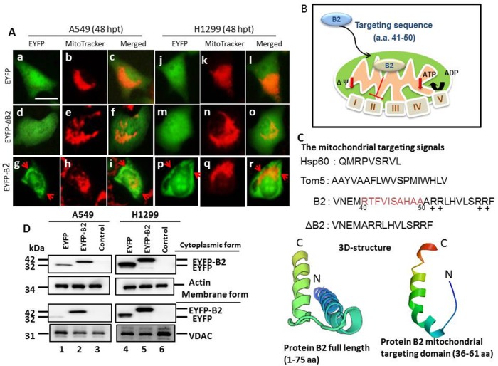
Figure 1. MitoTracker staining indicates the RGNNV B2 protein targets the mitochondria in human lung cancer cells.
Analysis of mitochondrial targeting of the EYFP-B2 fusion protein at 48 h post-transfection indicated yellow fluorescence in ∼4-5% of A549 cells (A: g-i and d; indicated by arrows) and H1299 cells (A: p-r) relative to cells with EYFP (A: a-c, in A549 cells; A: j-l, in H1299 cells) and EYFP-ΔB2 (del41RTFVISAHAA50) (A: d-f, in A549 cells; A: m-o, in H1299 cells). Phase-contrast images of EYFP-B2 transfected cells at 36 h post-transfection shows that the EYFP-B2 fusion protein targets mitochondria (indicated by arrows; A: i in A549 cells; A: r in H1299 cells). Scale bar: 10 μm. (B) RGNNV B2 protein construct used for mitochondrial targeting. (C) Various constructs of wild type and mutant forms of the RGNNV B2 protein used to identify the mitochondrial targeting sequence. The 3D-structure of full length of RGNNV protein B2 (1-75 aa) and B2 mitochondria targeting domain (36-61 aa) alone (see Materials and Methods) were shown, and that alpha helix also existing. N: N terminus; C: C terminus. (D) Immunoblotting using monoclonal antibodies against EYFP shows the protein distribution in mitochondrial and cytosolic fractions at 48 h post-transfection. The internal controls were actin (cytosolic fraction) and VDAC (mitochondrial membrane fraction). EYFP alone (negative control; lanes 1 and 4); EYFP-B2 (lanes 2 and 5), controls without vector (A549 and H1299 cells; lanes 3 and 6).

