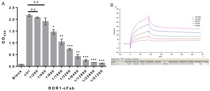
Figure 2. Confirmation of ROR1-cFab specificity and selectivity.
(A) ELISA. A 96-well plate was pre-coated with recombinant human ROR1 protein at a concentration of 50 ng/well. Serial dilutions of the ROR1-cFab were used as the first antibody for ELISA and the HRP-conjugated goat anti-human antibody (Fab specific) was used as the secondary antibody. Commercial anti-ROR1 antibody was used as a positive control (Ctrl). The absorbance was read at 450 nm after color development. (B) Surface Plasmon resonance (SPR) analysis. The ROR1 protein was diluted to 30 μg/mL in dilution buffer and then reacted in a running buffer containing serial concentrations of ROR1-cFab. Results were analyzed by the Biacore X100 software. The experiments were in triplicate and repeated at least twice. Data are shown as mean ± SD (n = 2, NS, not significant, *p < 0.05, **p < 0.01, and ***p < 0.001 compared with the control).

