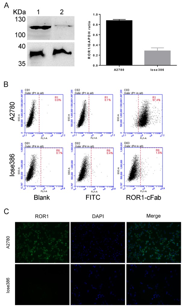
Figure 3. Assessment of the ROR1-cFab specificity in ovarian cancer cells.

(A) Western blot. It detected the level of ROR1 expression in ovarian cancer cells. Lane 1: supernatant of A2780 cell lysate; Lane 2: supernatant of Iose386 cell lysate. (B) Flow cytometry. Ovarian cancer A2780 and Iose386 cells were treated with or without ROR1-cFab for 1h and then incubated with a goat anti-human IgG (Fab specific)-FITC antibody for 1 h in the dark. The cells were then subjected to flow cytometry analysis of fluorescence intensity. (C) Immunofluorescence staining of ROR1. Ovarian cancer cells (A2780 and Iose386) were first grown and immunostained with ROR1-cFab. The experiments were in triplicate and repeated at least once.
