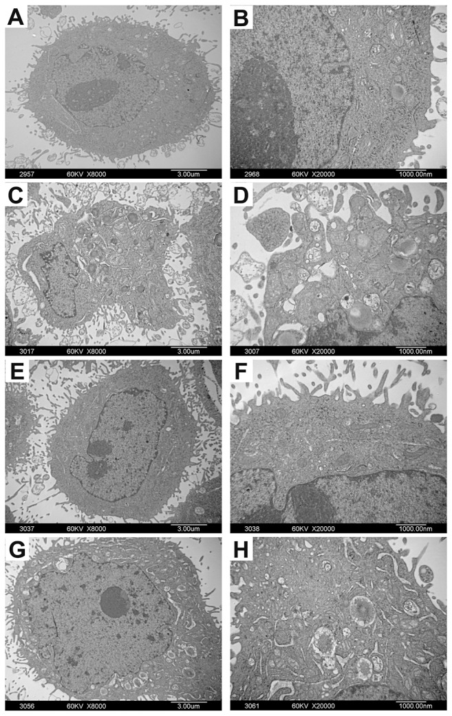
Figure 5. The ultrastructural changes of Bel-7402 and Hela cells treated with adenine.

Bel-7402 and Hela cells were treated with adenine (0.5mg/ml) for 48 hours. Figure (A, B) are control cells without adenine treatment. Figure (C-H) are cells treated with adenine. Under TEM, the cells treated with adenine show increase of nuclear heterochromatin, shrinkage and condensation of the nuclear membrane edge, irregular shaped nucleus and nuclear fragmentation (C, D); Increase of Microvilli on the cell surface (E, F); Increase of cytoplasmic vacuolization, mitochondria swelling and vacuolar degeneration. Rough endoplasmic reticulum became thickened and widened and ribosome de-granulated (G, H).
