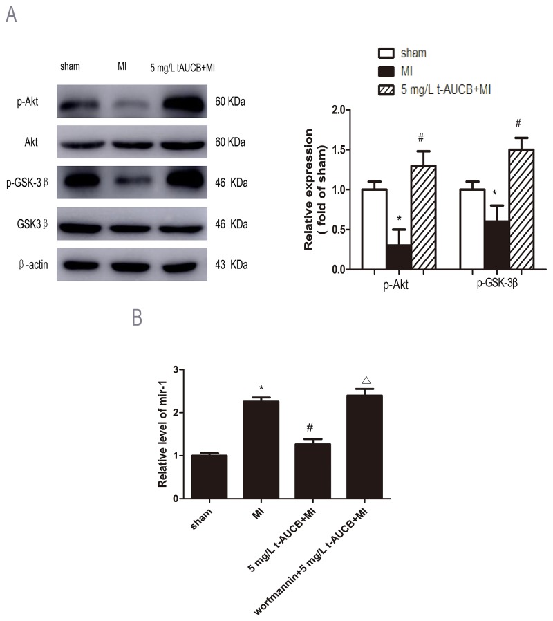
Figure 5. AKT/GSK3β signaling pathway participated in regulation of miR-1 by sEHi.
(A) Ischemic downregulated PKA and GSK3β expression in MI hearts, while 5 mg/L t-AUCB restored PKA and GSK3β expression. Measurements were made 24 h after MI. Left, examples of western blot bands; Right, relative expression level of p-Akt and p-GSK3β ratio to total AKT and GSK3β, respectively. Quantitation as mean ± SEM. *P<0.05 vs. sham group; #P<0.05 vs. MI group; n=3. (B) Levels of miR-1 was reduced in MI mice treated with 5 mg/L t-AUCB, while PI3K inhibitor wortmannin suppressed the downregulation of miR-1. miR-1 level were quantificated by real-time PCR with RNA samples isolated from mice hearts 24 h after MI. Data were expressed as mean ± SEM; *P<0.05 vs. sham group; #P<0.05 vs. MI group; ΔP<0.05 vs 5 mg/L t-AUCB +MI group, n=3.

