. 2017 Nov 29;6:e31558. doi: 10.7554/eLife.31558

Figure 6. Mutagenesis results support RNA-binding roles of regions Z1, Z3 and Z4 in HDX data, and key residues are identified.

(A) Identification of key residues in Z1. Positions of F32, R34, D36 and K39 in N-terminal helix of EZH2 are indicated in the ac/hsPRC2 crystal structure. K39A point mutant, triple mutant [F32A D36A K39A] and quadruple mutant [F32A R34A D36A K39A] were purified and tested for binding with (GGAA)10 RNA, together with the wild type 3 m hsPRC2. All proteins were used at successive threefold dilutions starting at 2.5 µM concentration. (B) Mutagenesis in Z2, Z3 and Z4 leads to identification of important residues in Z3 and Z4. All proteins were used at successive threefold dilutions starting at 5 µM concentration, and Kdapp values and errors are mean and standard derivation of three binding experiments performed at different days. (C) Histone methyltransferase activity assays for RNA-binding mutants, normalized to that of wild type and error calculated from three independent experiments. Red arrows indicate the mutants that disrupt RNA binding while maintaining normal catalytic activity. (D) Combining identified mutations into a single mutant [F32A D36A K39A and PRKKKR489-494NAAIRS] causes more severe binding defect for (GGAA)10 RNA. Both proteins were used at successive threefold dilutions starting at 5 µM concentration, and Kdapp values and errors are mean and standard derivation of three independent experiments.
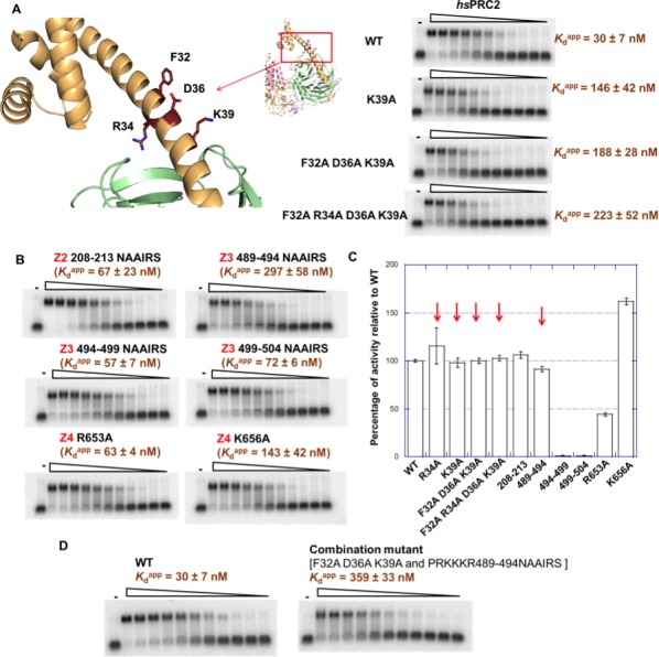
Figure 6—source data 1. Raw data in fRIP experiment.
DOI: 10.7554/eLife.31558.014

Figure 6.

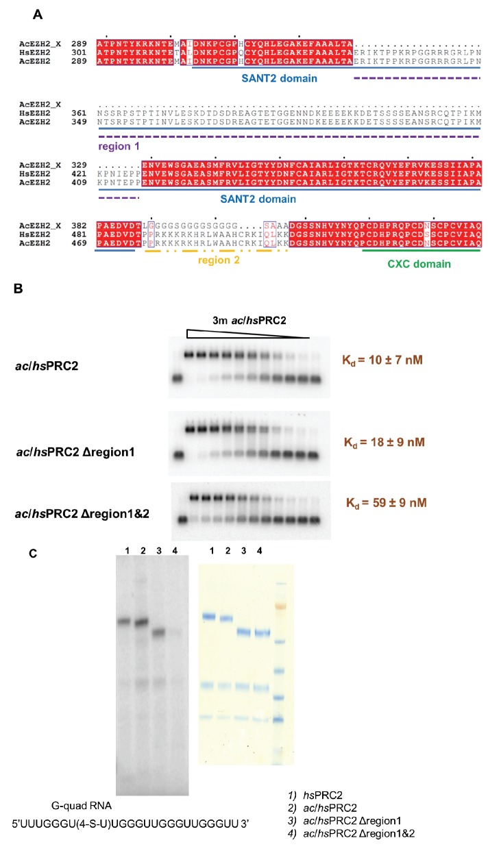
Figure 6—figure supplement 1. A basic region (adjacent to and include Z3 in HDX-MS result) of EZH2 is important for RNA binding.

Figure 6—figure supplement 1.

(A) Two previously deleted regions (region 1 and 2) in the ac/hsPRC2 crystal structure are indicated with dashed underlines. (Brooun et al., 2016). (B) ac/hsPRC2 wild type and two mutants (Δregion 1 and Δregion1 and 2 < crystallization construct>) were tested with (GGAA)10 RNA. Kdapp values and errors are mean and standard derivation of three independent experiments. (C) Protein-RNA crosslinking experiment using a 5’-32P-labeled G-quadruplex RNA (UUUGGGU(4-thio-U)UGGGUUGGGUUGGGUU). [Left panel] Protein samples and radiolabeled RNA were crosslinked using 365 nM UV light, resolved using SDS-PAGE and exposed to a phosphorimaging plate. Radiolabeled bands indicate that RNA is crosslinked to these polypeptides. [Right panel] Coomassie staining of the four protein samples on a SDS-PAGE gel.
Figure 6—figure supplement 2. Mutations in the context of the 5 m hsPRC2 (holoenzyme) exhibit binding defect specifically for RNA relative to DNA.
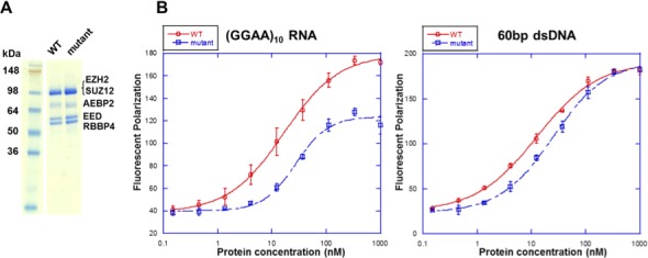
Figure 6—figure supplement 2.

(A) Wild type and mutant [F32A R34A D36A K39A PRKKKR489-494NAAIRS] EZH2 were expressed and purified in the context of the 5 m hsPRC2 (EZH2-SUZ12-EED-RBBP4-AEBP2). The recombinant 5 m hsPRC2 complexes reconstituted with the baculovirus system were resolved using a Novex 10–20% Tris-Glycine gel (WedgeWell format, ThermoFisher) and stained with Coomassie Blue for visualization. Individual polypeptides are marked. (B) Wild type and mutant 5 m hsPRC2 were tested for binding with a (GGAA)10 RNA and a 60 bp double-stranded DNA (produced by annealing two single-stranded (GC)30 DNA oligos) using fluorescent anisotropy. 5 nM of the Alexa488 fluorophore-attached RNA or DNA were folded and incubated as described for the EMSA experiments. RNA binding was measured at high-salt condition (100 mM KCl and 2.5 mM MgCl2) as used in the EMSA experiments, and DNA binding was measure in low-salt condition (10 mM KCl and 0.25 mM MgCl2) to enhance the binding. All proteins were used at successive 3-fold dilutions starting at 1 µM concentration, and all polarization values and errors are mean and standard derivation of three binding experiments performed on different days.
Figure 6—figure supplement 3. Identified residues are important for (GGAA)10 RNA interaction in vivo.
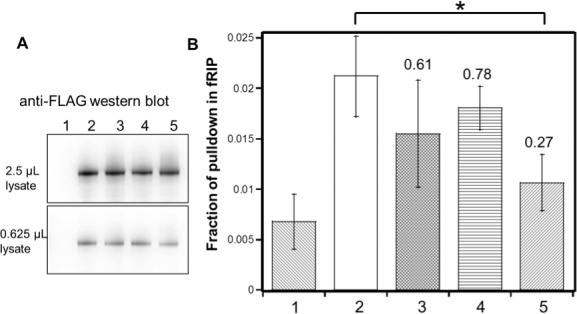
Figure 6—figure supplement 3.

FLAG-tagged hsEZH2 (wild type or mutants) was co-expressed with (GGAA)10-MS2 RNA in HEK293T cells, and formaldehyde RNA immunoprecipitation (fRIP) was performed to evaluate the percentage of pull down of the RNA by each hsEZH2 variant. hsEZH2 variant used in each lane is: 1. no EZH2 (negative control); 2. wild type; 3. [K39A]; 4. [F32A R34A D36A K39A]; 5. [F32A R34A D36A K39A and PRKKKR489-494NAAIRS]. (A) Western Blot using anti-FLAG antibody was performed to compare the expression levels of exogenous hsEZH2 wild type and mutants. Two different amounts (2.5 or 0.625 µL) of lysate were used to better evaluate the expression levels. (B) Fraction of pulldown of (GGAA)10 RNA by each hsEZH2 variant is plotted in the bar graph, and the mean ± S.D. of three independent experiments is shown. *p-value<0.05. The number above each of the three mutants is the fold change of RNA pulldown relative to wild type with background reduction by subtracting the negative control value.