. 2017 Nov 13;6:e29176. doi: 10.7554/eLife.29176

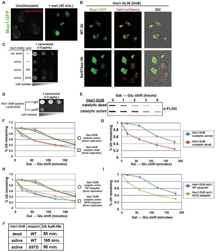
Figure 5. Ser57Asp phosphomimetic ubiquitin bypasses an artificial DUB checkpoint.

(A) Analysis of Mup1-GFP (green) localization in wildtype yeast cells (strain SEY6210) grown in minimal media lacking methionine (left) or stimulated with methionine for 45 min (right). Cells are stably expressing Vph1-mCherry (red), a marker of the vacuolar membrane. (B) Mup1-GFP localization in yeast cells (strain SEY6210) where the endogenous Hse1 locus has been fused to the UL36 deubiquitylating enzyme, encoding an Hse1-UL36 fusion protein. This creates an artificial DUB checkpoint at ESCRT-0. The top panel of cells are expressing wildtype ubiquitin, whereas the bottom panel of cells are expressing Ser57Asp phosphomimetic ubiquitin. As in (A), cells are stably expressing Vph1-mCherry (red), a marker of the vacuolar membrane. (C and D) Analysis of yeast growth in the presence of canavanine. (E) Slot blot analysis of FLAG-ubiquitin levels following a galactose induction/glucose repression. (F–J) Quantitation of slot blot experiments (as shown in E, n = 3) from yeast cells expressing catalytic active or catalytic dead Hse1-UL36 fusion (F and G) or catalytic active Hse1-UL36 in the presence of wildtype or Ser57Asp phosphomimetic ubiquitin (H and I). Data were averaged over multiple experiments (G and I) (n = 3, error bars indicate standard deviation) and trend line regression was used to calculate ubiquitin half life (J).

Figure 5.

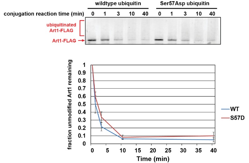
Figure 5—figure supplement 1. Ser57Asp phosphomimetic ubiquitin does not alter the rate of Rsp5-mediated conjugation in vitro.

Figure 5—figure supplement 1.

In this experiment, recombinant yeast proteins GST-Uba1 (E1), 6xHIS-Ubc1 (E2), 6xHIS-Rsp5 (E3), Art1-FLAG (substrate), and ubiquitin or Ser57Asp phosphomimetic ubiquitin were incubated at room temperature in the presence of ATP. Each conjugation time course was analyzed by α-FLAG immunoblotting and quantified over three independent experiments (n = 3).
Figure 5—figure supplement 2. Ser57Asp phosphomimetic mutation does not alter binding of ubiquitin to ESCRT-I or ESCRT-II in vitro.

Figure 5—figure supplement 2.

In this experiment, recombinant ESCRT-I and ESCRT-II complexes were used as bait in the capture of wild-type or Ser57Asp phosphomimetic ubiquitin. Captured samples were analyzed by SDS-PAGE followed by immunoblotting for total ubiquitin.
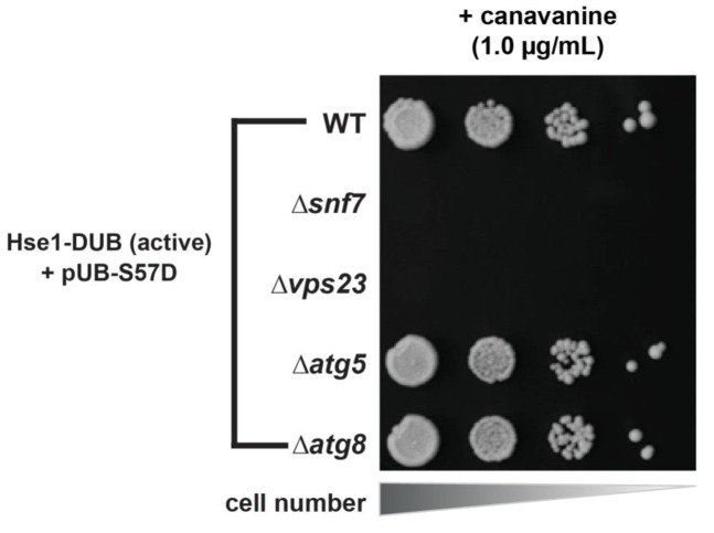
Figure 5—figure supplement 3. DUB bypass by Ser57Asp phosphomimetic ubiquitin requires an in-tact ESCRT pathway while the autophagy pathway is dispensible.

Figure 5—figure supplement 3.

Yeast strains (SEY6210 background) expressing Hse1-UL36 fusions and Ser57Asp phosphomimetic ubiquitin were analyzed for growth in the presence of canavanine in order to determine the genetic requirements for the canavanine resistance phenotype.
Figure 5—figure supplement 4. Bypass of a Can1-DUB fusion by Ser57Asp phosphomimetic ubiquitin.
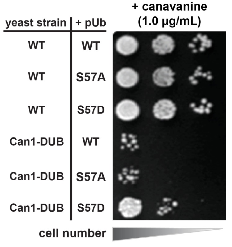
Figure 5—figure supplement 4.

Wild-type yeast strains (SUB280 background) or an isogenic strain encoding an endogenous Can1-UL36 DUB fusion were analyzed for canavanine sensitivity/resistance in the presence of wild-type, Ser57Ala, or Ser57Asp phosphomimetic ubiquitin.
Figure 5—figure supplement 5. Phenotypic analysis of yeast cells expressing Leu73Pro ubiquitin.
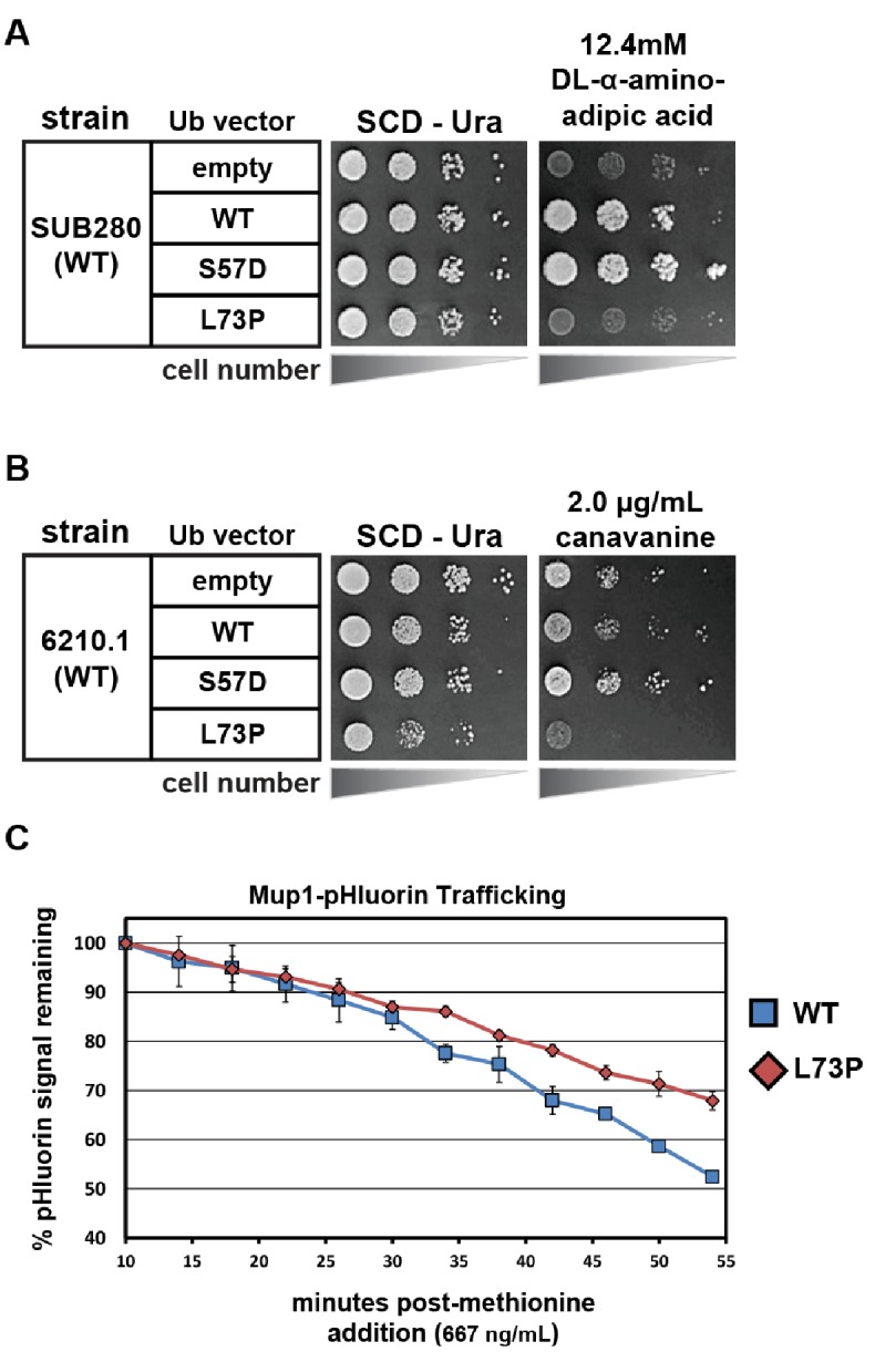
Figure 5—figure supplement 5.

(A) SUB280 yeast cells (expressing ubiquitin from a single pLYS source plasmid) were transformed with a second ubiquitin-expressing plasmid (pURA) and grown in the presence of DL-α-aminoadipic acid to counterselect against the pLYS plasmid (‘plasmid shuffle’). (B) Wildtype yeast cells (SEY6210.1 background) transformed with either empty vector or plasmids expressing ubiquitin (wildtype, S57A or L73P) from the ADH1 promoter were plated on minimal media with or without canavanine. (C) Wildtype yeast cells expressing pHluorin-tagged Mup1 (SEY6210.1 background) transformed with either empty vector or plasmids expressing ubiquitin (wildtype, S57A or L73P) from the ADH1 promoter were monitored using flow cytometry measurements of pHluorin signal in a time course following methionine stimulation.